
A crowd presses around an opened body. The anatomist stands beside it, while a skeleton rises above the gathering. This is the frontispiece to Andreas Vesalius’s De humani corporis fabrica, published in 1543. Before a reader reaches an anatomical description, the book presents a claim about how knowledge should be made: the author belongs at the body, performing and explaining the work. The scene is carefully composed, not a candid record of one crowded lesson.
The Fabrica—usually translated as “On the Fabric of the Human Body”—became an influential anatomical publication. Its importance lies in the relationship between what Vesalius examined and what readers could examine on the page. Dissection could challenge an inherited account, but a book needed to make that challenge legible to someone who had never stood beside the same body. Text, figures and the author’s self-presentation helped bridge that distance.
PLACE & TIME
Basel and Padua · 1543–1555 CE
Basel and Padua
Publication in Basel; anatomical teaching in northern Italy
1543–1555 CE
The map marks Basel as a publishing centre, not the location of every dissection represented in the book.
Bodies were being studied before the Fabrica
Vesalius worked within a long medical tradition. Galen, a Greek physician active in the Roman Empire in the second century, remained a major authority on bodily structure and function. Much of his anatomical work depended on animals. That created problems when descriptions were applied to human bodies. Yet Renaissance anatomists did not discover dissection after centuries in which nobody had looked: the National Library of Medicine’s timeline includes medieval dissection teaching and manuals.
The relationship with Galen was also more complicated than simple rebellion. Vesalius learned from him, retained much of his account and used close anatomical investigation to identify disagreements. The NLM’s historical description emphasises both reliance and criticism. An authority could provide the questions and vocabulary through which someone eventually found a reason to correct it. There was no clean division between people who read books and people who examined bodies.
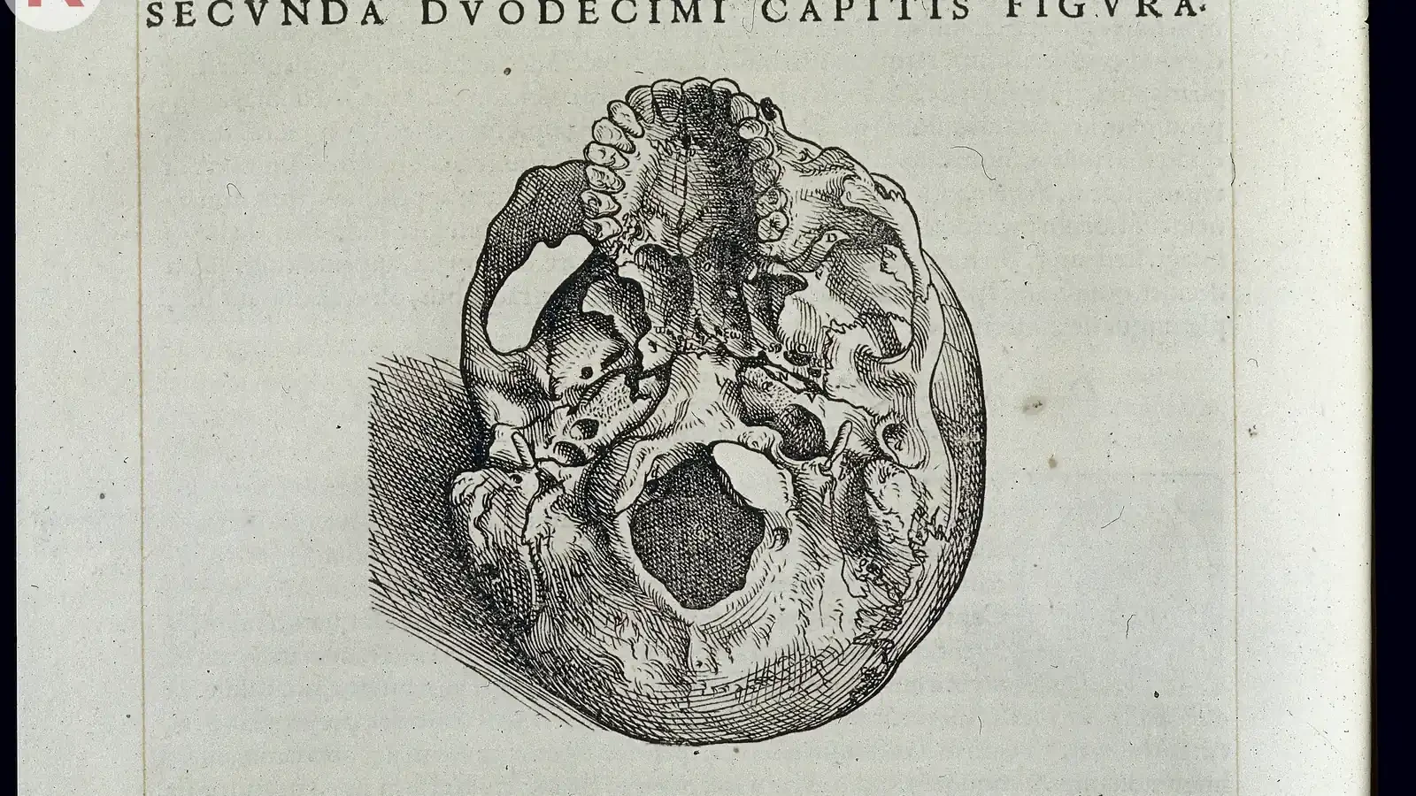
The book’s structure lets us follow that work at the level of a particular claim. Book I, chapter 10 treats the lower jaw, including the assertion that the human jaw is essentially a single bone. The modern translation’s contents identify the passage and its original pages. A reader can move from a general reputation for anatomical innovation to a specific part, illustration and argument that can be inspected. The question becomes what the description says about this structure and how it supports that account.
TURNING POINTS
A book within a developing investigation
- 1538
Tabulae anatomicae sex
Vesalius publishes six anatomical tables; the later Fabrica was not his first effort to communicate anatomy visually.
- 1543
Fabrica and Epitome
The substantial seven-book work and a shorter companion appear in Basel, printed by Johannes Oporinus.
- 1555
A revised Fabrica
A second edition makes the work itself part of an ongoing process of correction and revision.
Publication dates orient the object’s history. They do not identify the beginning of human dissection or a moment when anatomical knowledge became complete.
The opening image puts manual work at the centre
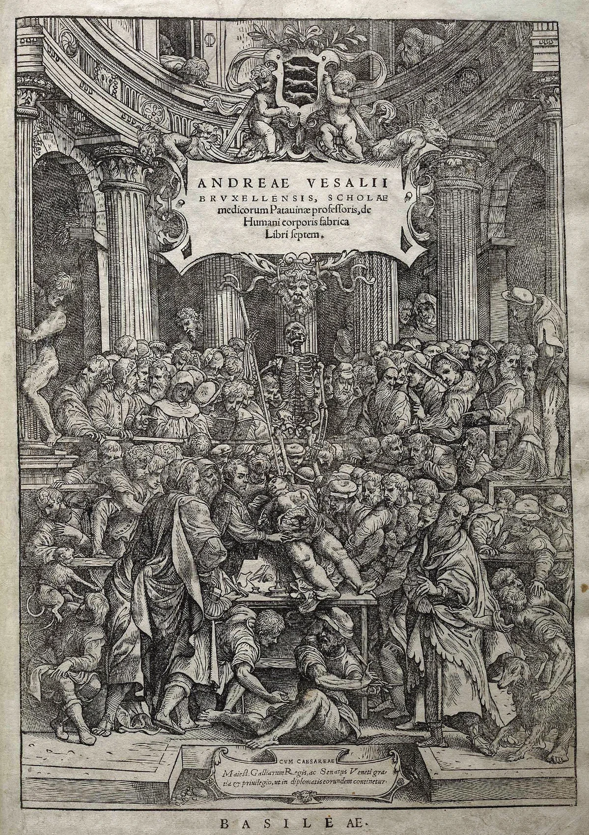
The frontispiece rearranges authority. Vesalius is not merely a learned figure separated from the operation while another person handles the body. He is placed at the dissection itself. The crowd supplies an audience, and the skeleton supplies another object of attention. These are visual choices with a purpose. The image asks a reader to associate the author’s authority with direct bodily work, not simply with his ability to repeat a text.
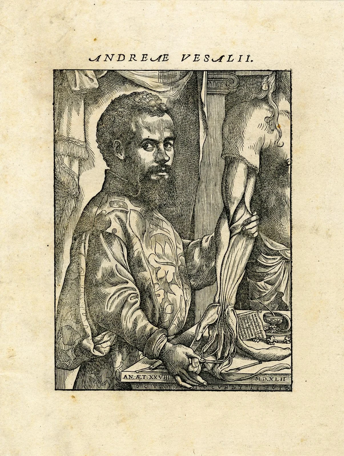
A second image makes that message more personal. The author’s portrait shows him handling a dissected arm while looking towards the viewer. The pose links identification—this is the man named on the book—to a claim of competence. It is still a portrait, composed to make someone recognisable and persuasive. Its value as evidence lies partly in what the author wanted his audience to understand about him.
KEEP THE DISTINCTION
Two images, two kinds of authority
The crowded dissection
An anatomical programme centred on the author’s presence at the body and before an audience.
That this exact gathering took place or that every spectator accepted the same conclusions.
The author with an arm
A public identity combining learned authorship with manual anatomical skill.
The circumstances of every dissection or the accuracy of every description elsewhere in the book.
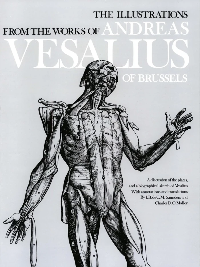
A labelled anatomical figure
A selected structure organised for examination and connection with the text.
That the represented body was typical in every respect or that no interpretive choices shaped the drawing.
The images also expose a silence. The opened body is central to the demonstration, but the person whose body it was does not speak through the frontispiece. That does not allow us to invent a biography or claim a particular history of consent from the image alone. It does require us to notice the uneven distribution of visibility: author and observers acquire a place in the story, while the subject becomes material through which others make knowledge.
A figure could organise what a body made difficult to see
A dissection unfolds through time. Parts are exposed, moved or removed, and the view changes as work proceeds. A printed figure can select one stage, adopt a useful angle and place labels where readers can follow them. That gives an anatomical book an advantage beyond preserving a moment. It can organise bodily complexity into a sequence that can be revisited, compared and discussed.
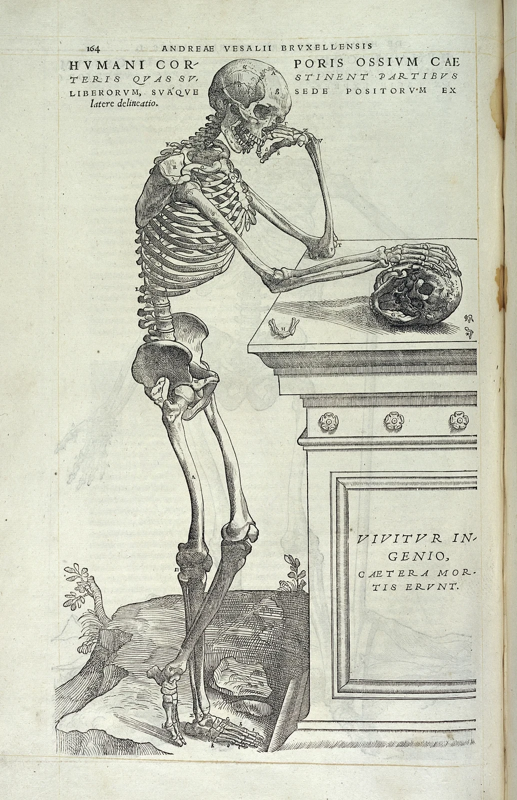
The skeleton leaning towards a skull offers a particularly clear example. Its bones are displayed, but its pose also turns anatomy into a reflective scene. The figure is neither a neutral inventory nor simply decoration. A memorable arrangement helps the structure remain available for study. Asking how the illustration works is more productive than deciding that it must be either accurate science or expressive art.
Labels also change how a page is used. They let a reader move between a visible part and a written explanation. Looking at the figure without consulting the text can miss a qualification or a distinction; reading the text without the figure can make a spatial relationship harder to follow. The Fabrica offers its argument through this movement between formats. A distant reader is given something to do, rather than only a conclusion to accept.
KEY IDEA
Selecting and arranging a view can make a bodily relationship easier to examine.
The author’s name does not identify every maker
The book appeared in Basel from the workshop of Johannes Oporinus, while Vesalius’s anatomical teaching was associated with Padua. Images had to pass through drawing, cutting and printing before they reached a reader. Cambridge’s exhibition describes that movement across places and skills. The work’s achievement cannot be reduced to one person observing something and a printer mechanically copying it.
The attribution of the illustrations makes the point more sharply. Jan Stephan van Calcar is associated with Vesalius’s earlier anatomical tables and with the author’s portrait. Older accounts also treated him as the sole artist behind the Fabrica. The NLM notes that subsequent scholarship identifies the work of several artists. We should preserve that uncertainty rather than attach a confidently named hand to every figure.
LOOK FROM HERE
What had to work together?
Dissection supplied opportunities to compare a written description with bodily structures, while also creating practical problems of access and visibility.
Artists and block cutters turned selected structures into reproducible figures. Their skills helped determine what remained clear on the printed page.
Readers used text and images together. A page made a view portable without giving every reader the same practical experience as the anatomist.
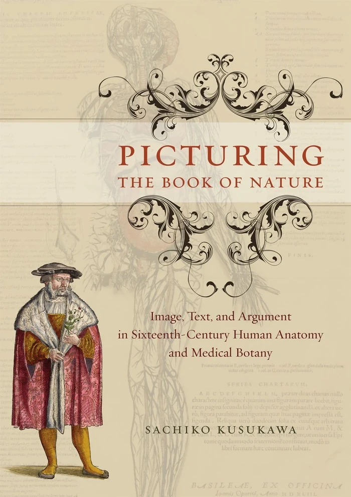
Sachiko Kusukawa’s study of visual argument places anatomy alongside medical botany: pictures did not merely accompany learned writing, but could help conduct an argument. That is a useful question to bring back to the Fabrica. Instead of praising its beauty and moving on, ask what a particular illustration helps a reader distinguish, compare or understand. Its appearance is part of its intellectual work.
A published page did not finish the investigation
The Fabrica was a substantial seven-book work, while the shorter Epitome, also published in 1543, offered another route into its anatomical programme. Wellcome’s copy documents this companion publication. Their coexistence matters: communicating anatomy was not one task for one audience. Scale, organisation and the amount of explanation shaped how different books could be used.
Revision matters too. A second edition appeared in 1555, and surviving annotations have become evidence for the work’s further development. Research on Vesalius’s annotations shows why a celebrated first edition should not be treated as the last word. The claim that bodies could correct books had a consequence for his own book as well. Printed authority could be influential and still remain open to alteration.
TAKE THIS WITH YOU
The Fabrica made anatomy available for inspection
Keep the tradition in view
Galen shaped Vesalius’s questions as well as supplying claims he challenged; dissection already had a history.
Read between image and text
Labels, viewpoints and sequences organise evidence. A vivid anatomical figure is not a transparent substitute for a body.
Look beyond the author
Artists, printers, readers and the people whose bodies were examined belong within the history of anatomical knowledge.
A book can invite scrutiny and still become the subject of an official judgement about what it is permitted to say. The next chapter follows Galileo’s Dialogue into the proceedings of 1633. There, the question of how to argue from evidence met the question of who had authority to permit an argument at all.


.jpg)
